
This article briefly describes the structure, functions, and metabolic significance of adipose tissue, with particular emphasis on the storage, mobilization, and regulation of fatty acids. Adipose tissue is a specialized connective tissue that plays a fundamental role in maintaining the body’s energy balance. It stores excess energy in the form of triacylglycerols and releases fatty acids to meet metabolic demands during periods of energy deficiency. Beyond its role in lipid storage, adipose tissue also functions as an active endocrine organ and contributes to thermoregulation.
Organization and Functions of Adipose Tissue
Adipose tissue is a specialized connective tissue that functions as the body’s principal site for the storage of metabolic energy in the form of triglycerides and serves as a major source of fatty acids during periods of increased energy demand. In addition to storing lipids, it provides mechanical protection, thermal insulation, and endocrine functions that help regulate whole-body metabolism. Based on its structural and physiological characteristics, adipose tissue is classified into two major types, white adipose tissue (WAT) and brown adipose tissue (BAT).
White adipose tissue is the predominant form and is widely distributed beneath the skin, around major blood vessels, and within the abdominal cavity. Its adipocytes are large, spherical cells containing a single lipid droplet that occupies most of the cellular volume, displacing the nucleus and mitochondria toward the cell periphery (Figure 1). This lipid droplet primarily stores triglycerides and sterol esters and is surrounded by a phospholipid monolayer with its hydrophilic head groups oriented toward the cytosol.
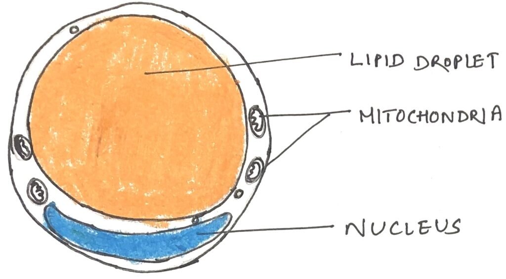
The surface of the lipid droplet also contains specialized proteins, including perilipin, together with enzymes that regulate the synthesis and degradation of triacylglycerols, thereby controlling lipid storage and mobilization. In healthy young adults, white adipose tissue accounts for approximately 15% of total body mass. Rather than serving as a passive fat depot, adipocytes are highly metabolically active cells that respond rapidly to hormonal signals and coordinate lipid metabolism through continuous communication with the liver, skeletal muscles, and heart.
Fatty Acid Storage and Lipid Metabolism in Adipocytes
Adipocytes are highly active metabolic cells that participate in several essential biochemical pathways involved in energy metabolism. Similar to many other cell types, they generate ATP through glycolysis, the citric acid cycle, and oxidative phosphorylation while also oxidizing fatty acids to meet cellular energy demands. Following carbohydrate-rich meals, excess glucose can be converted into fatty acids through de novo lipogenesis, although the liver serves as the primary site of this process in humans. These newly synthesized fatty acids are subsequently esterified to form triacylglycerols, which accumulate within large intracellular lipid droplets as long-term energy reserves.
In addition to producing fatty acids from glucose, adipocytes efficiently take up circulating lipids supplied by the liver in the form of very-low-density lipoproteins (VLDLs) and by the intestine as chylomicrons, particularly after fat-rich meals. By integrating these metabolic processes, adipose tissue functions as the body’s principal reservoir for storing excess energy in the form of triacylglycerols.
Mobilization of Stored Fatty Acids
During periods of increased energy demand, such as between meals or during fasting, adipocytes begin to mobilize their stored triacylglycerols. Lipolytic enzymes break down triacylglycerols into free fatty acids and glycerol. The released fatty acids enter the bloodstream and are transported to energy-demanding tissues. Skeletal muscle and the heart use these fatty acids as important sources of fuel. Epinephrine strongly stimulates this process by activating a cyclic AMP (cAMP)-dependent signaling pathway. This pathway phosphorylates perilipin, the protein that surrounds lipid droplets. Phosphorylated perilipin allows lipases that act on triacylglycerols, diacylglycerols, and monoacylglycerols to reach the stored lipids more efficiently. Hormone-sensitive lipase is also activated by phosphorylation. However, the activation of perilipin plays a major role in increasing lipolysis. In contrast, insulin suppresses lipolysis by reducing lipase activity. As a result, insulin promotes the storage of triacylglycerols and helps maintain the body’s energy reserves.
Triacylglycerol Recycling and Endocrine Functions of Adipose Tissue
Adipose tissue continuously balances the breakdown and synthesis of triacylglycerols through a process known as the triacylglycerol substrate cycle. During lipolysis, adipocytes release large amounts of free fatty acids. However, not all of these fatty acids leave the cell. A substantial proportion is rapidly re-esterified and converted back into triacylglycerols, thereby replenishing intracellular lipid stores.
Unlike many other tissues, adipocytes cannot directly recycle the glycerol released during lipolysis because they lack the enzyme glycerol kinase. Instead, they generate glycerol-3-phosphate from pyruvate through a metabolic pathway called glyceroneogenesis. This pathway depends on the activity of the cytosolic enzyme phosphoenolpyruvate carboxykinase (PEPCK) and provides the glycerol backbone required for the resynthesis of triacylglycerols.
Beyond serving as the body’s principal lipid reservoir, adipose tissue also functions as an important endocrine organ. It synthesizes and secretes a variety of signaling molecules, known as adipokines, that communicate the status of energy stores to other organs. These hormones regulate lipid and carbohydrate metabolism, helping coordinate energy balance throughout the body.
Brown Adipose Tissue and Heat Production
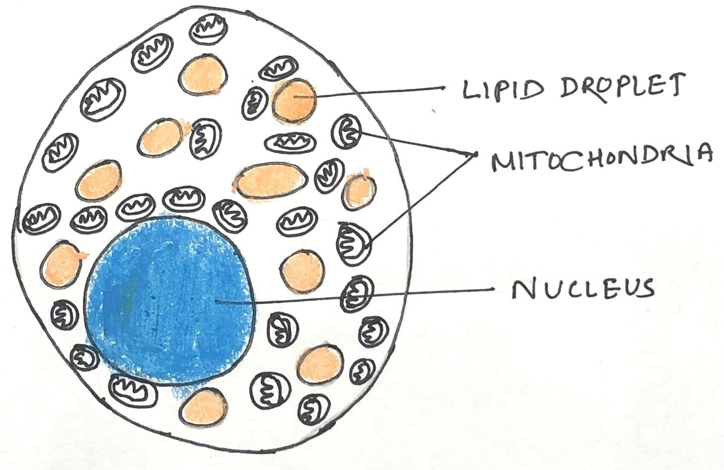
Brown adipose tissue (BAT) is abundant in small mammals, hibernating animals, and newborn humans. It plays a crucial role in maintaining body temperature. Unlike white adipose tissue (WAT), which primarily stores energy, BAT is specialized for heat generation. Brown adipocytes are smaller than white adipocytes and contain numerous small lipid droplets instead of a single large one (Figure 2). They also possess a large number of mitochondria and are richly supplied with blood vessels and nerve fibers. The high mitochondrial content, together with the haemoglobin present in the dense capillary network, gives BAT its characteristic brown appearance.
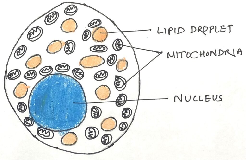
The most distinctive feature of brown adipocytes is the presence of uncoupling protein 1 (UCP1), also known as thermogenin. This protein enables BAT to produce heat through a process called non-shivering thermogenesis. During this process, fatty acids stored within lipid droplets are released and transported into the mitochondria. They are completely oxidized through β-oxidation and the citric acid cycle, generating NADH and FADH₂. These reduced coenzymes donate electrons to the electron transport chain, which pumps protons across the inner mitochondrial membrane.
In white adipose tissue, these protons return to the mitochondrial matrix through ATP synthase, driving ATP production. In contrast, brown adipose tissue uses UCP1 to provide an alternative pathway for proton re-entry. This bypasses ATP synthase and prevents ATP synthesis. Instead, the energy stored in the proton gradient is released as heat. This mechanism helps maintain body temperature, especially during exposure to cold environments.
Development and Physiological Significance of Brown and Beige Adipose Tissue
The formation of brown adipose tissue begins during fetal development. Around the twentieth week of gestation, precursor fibroblast-like cells differentiate into brown adipocytes. At birth, BAT constitutes approximately 1–5% of an infant’s body mass. It is strategically distributed around major blood vessels and vital organs, including the kidneys, pancreas, adrenal glands, and the large blood vessels supplying the brain and abdomen. This location enables the heat generated by BAT to protect essential organs from cold stress immediately after birth.
As growth continues, the amount of brown adipose tissue gradually declines while white adipose tissue becomes the predominant fat depot. In healthy adults, BAT accounts for only a small fraction of total adipose tissue, representing less than 0.1% of body weight. Despite this reduction, adults retain a population of specialized adipocytes within white adipose tissue that can acquire characteristics similar to brown adipocytes. These cells, known as beige adipocytes, develop in response to prolonged cold exposure or stimulation of β-adrenergic receptors.
Beige Adipose Tissue and Metabolic Adaptation
Beige adipocytes contain multiple small lipid droplets, possess abundant mitochondria, and express uncoupling protein 1 (UCP1). Like brown adipocytes, they generate heat through non-shivering thermogenesis. In addition to oxidizing their own stored fatty acids, these cells actively remove fatty acids and glucose from the bloodstream to support the production of heat. Their high rate of glucose utilization allows clinicians to detect metabolically active BAT using positron emission tomography (PET) scanning. Hormonal and genetic factors also regulate the formation and activity of brown and beige adipose tissue.
Individuals with pheochromocytoma, a tumor that causes excessive secretion of epinephrine and norepinephrine, often develop increased amounts of beige adipose tissue. The nuclear transcription factor PPARγ plays a key role in the differentiation of white, brown, and beige adipocytes. It also helps the body adapt to changes in environmental temperature. Another important regulator is irisin, a peptide hormone released by skeletal muscles during physical exercise. Irisin stimulates the formation of beige adipocytes. As a result, heat production and energy expenditure remain elevated even after exercise has ended.
Conclusion
Adipose tissue is far more than a passive reservoir of stored fat. It is a dynamic and metabolically active organ. It plays a central role in energy storage, fatty acid mobilization, and the regulation of whole-body metabolism. White adipose tissue stores excess energy in the form of triacylglycerols. When the body’s energy demand increases, it releases fatty acids into the bloodstream. In contrast, brown and beige adipose tissues generate heat through thermogenesis. This process helps maintain body temperature, especially during exposure to cold. In addition, adipose tissue functions as an endocrine organ by secreting hormones that regulate lipid and carbohydrate metabolism.
A deeper understanding of the structure, functions, and regulation of adipose tissue has greatly improved our knowledge of energy homeostasis and metabolism. This knowledge has revealed the diverse roles of adipose tissue in maintaining metabolic health. It has also provided valuable insights into the prevention, diagnosis, and management of metabolic disorders. Continued research in this field is expected to support the development of more effective therapeutic strategies in the future.
You may also like:
- Proteins Facilitate the Transbilayer Movement of a Lipid Molecule
- Maintenance of blood glucose level by the hormones insulin and glucagon
- The metabolism of fuel in fasting and starvation
- Cholesterol Transport and Lipoprotein Metabolism

I, Swagatika Sahu (author of this website), have done my master’s in Biotechnology. I have around fourteen years of experience in writing and believe that writing is a great way to share knowledge. I hope the articles on the website will help users in enhancing their intellect in Biotechnology.