
This article describes the structure, function, and types of hemoglobin, the molecular basis of sickle cell anemia, its clinical manifestations, and the evolutionary significance of the sickle cell trait. Hemoglobin is an essential globular protein in red blood cells that transports oxygen from the lungs to tissues throughout the body. Its structure and oxygen-binding properties are closely linked to its physiological function. Changes in the genes encoding globin chains can alter the properties of hemoglobin and lead to inherited disorders. Sickle cell anemia is a notable example in which a single amino acid substitution causes abnormal hemoglobin interactions and changes the shape and function of red blood cells.
Hemoglobin and Its Structure
Hemoglobin is an oxygen-binding protein present in red blood cells (erythrocytes), where it facilitates the transport of oxygen throughout the body. Mature human erythrocytes lack a nucleus and the cellular machinery required for division, and they generally circulate for approximately 120 days before being removed from the bloodstream. These cells contain a high concentration of hemoglobin dissolved in their cytoplasm. Under normal physiological conditions, hemoglobin in arterial blood is highly saturated with oxygen, whereas its oxygen saturation decreases in venous blood as oxygen is delivered to peripheral tissues.
Hemoglobin is a globular protein with an approximate diameter of 5.5 nm. It consists of four polypeptide subunits, each containing a heme prosthetic group that enables oxygen binding. Structural studies using X-ray crystallography have identified two principal conformational states of hemoglobin: the relaxed (R) state and the tense (T) state. Although oxygen can bind to both conformations, the R state exhibits a greater affinity for oxygen than the T state.
Oxygen Binding and Cooperative Behavior
Hemoglobin must efficiently capture oxygen in the lungs and release it in peripheral tissues. A protein with an excessively high oxygen affinity would retain oxygen and release insufficient amounts to the tissues. Conversely, a protein with a very low affinity would be unable to bind adequate oxygen in the lungs. Hemoglobin overcomes this limitation through changes in its three-dimensional structure during oxygen binding.
In the absence of oxygen, hemoglobin predominantly favours the low-affinity T state. As oxygen molecules bind to its subunits, conformational changes progressively favour the high-affinity R state. This transition increases the affinity of the remaining unoccupied binding sites for oxygen. Consequently, hemoglobin exhibits a characteristic sigmoidal oxygen-binding curve, reflecting its ability to bind oxygen cooperatively rather than independently.
Allosteric Regulation of Hemoglobin
Allosteric proteins are molecules whose binding properties at one site are influenced by the binding of a ligand or regulator at another site. This regulation occurs through changes in protein conformation that alter the activity or binding affinity of the protein. Depending on their effects, allosteric regulators may function as activators or inhibitors.
Allosteric interactions are classified as homotropic or heterotropic. In homotropic regulation, the ligand responsible for modifying the protein’s conformation is the same molecule that binds to its functional sites. In heterotropic regulation, the regulator is a different molecule from the primary ligand. Hemoglobin demonstrates homotropic allostery because oxygen acts both as the ligand and as a positive regulator of subsequent oxygen binding. The binding of one oxygen molecule increases the affinity of the remaining subunits for oxygen, providing the molecular basis for cooperative oxygen binding.
Types of Hemoglobin
Human hemoglobin occurs in several forms that differ in their globin-chain composition and physiological roles. The four important types are HbA, HbA₂, HbF, and HbS.
Hemoglobin A (HbA)
Hemoglobin A is the predominant form of hemoglobin in healthy adults, accounting for approximately 95–98% of total adult hemoglobin. It consists of two α-globin chains and two β-globin chains and is primarily responsible for oxygen transport in the body.
Hemoglobin A₂ (HbA₂)
HbA₂ is a minor adult hemoglobin that normally constitutes approximately 2–3.5% of total hemoglobin. It contains two α-globin chains and two δ-globin chains. Although it also transports oxygen, its contribution to total oxygen transport is relatively small.
Fetal Hemoglobin (HbF)
HbF is the predominant hemoglobin during fetal development and consists of two α-globin chains and two γ-globin chains. Its higher affinity for oxygen compared with adult hemoglobin facilitates oxygen transfer from maternal blood to the developing fetus across the placenta. HbF levels decrease after birth as adult hemoglobin production increases.
Hemoglobin S (HbS)
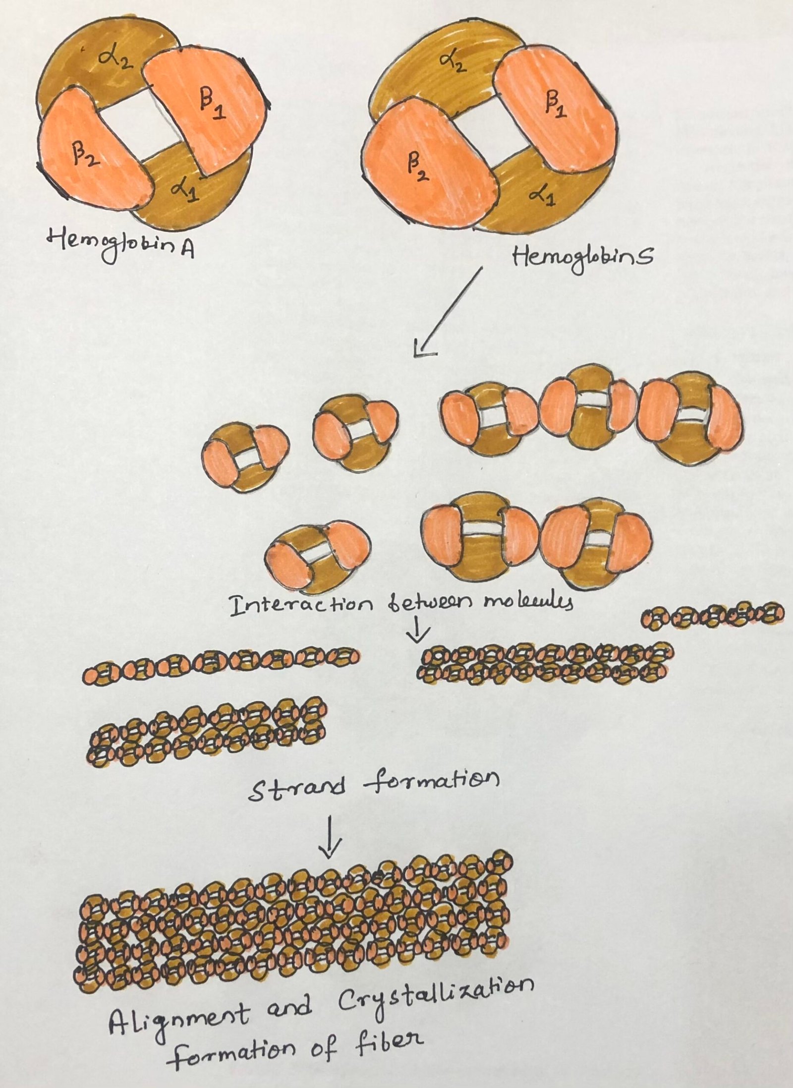
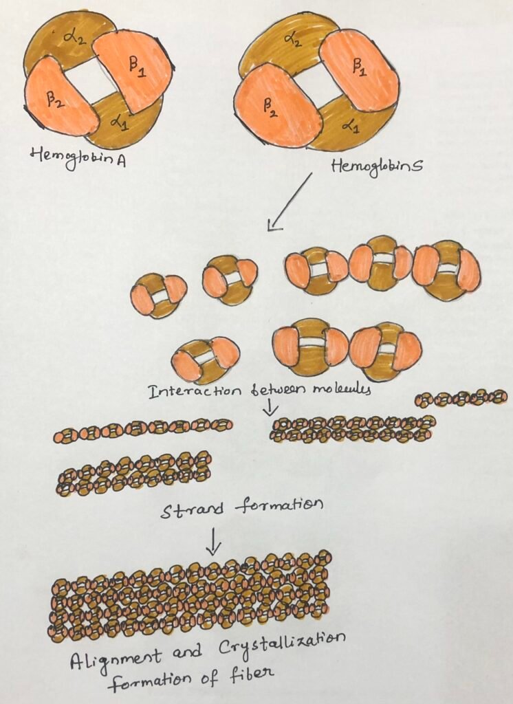
HbS is a structurally altered hemoglobin variant associated with sickle cell disease. It results from a mutation in the HBB gene that replaces glutamic acid with valine at the sixth amino acid position of the β-globin chain. Under low-oxygen conditions, HbS molecules can aggregate into long polymers, causing red blood cells to become rigid and sickle-shaped.
Molecular Basis of Sickle Cell Anemia
Hemoglobin exhibits considerable genetic diversity, with variations arising from changes in the genes encoding its globin chains. These changes may alter individual amino acids within the protein, sometimes affecting its structural properties and physiological function. The different forms of a gene are known as alleles. Since humans typically inherit one copy of each autosomal gene from each parent, the combination of alleles determines which hemoglobin variants an individual produces.
Sickle cell anemia is an inherited blood disorder that develops when an individual inherits two copies of the sickle hemoglobin allele, one from each parent. The condition is characterized by chronic anemia and the formation of abnormally shaped red blood cells. These cells become rigid and less flexible, which can interfere with circulation and reduce oxygen delivery to tissues.
Molecular Changes in Hemoglobin S and Red Blood Cell Sickling
The molecular defect responsible for this disorder involves a specific substitution in the β-globin chain of hemoglobin. In HbS, the sixth amino acid residue of the β-globin chain is valine instead of glutamic acid, as found in normal adult hemoglobin (HbA). Glutamic acid carries a negative charge at physiological pH, whereas valine is uncharged and hydrophobic. This single substitution changes the chemical character of the hemoglobin surface, creating a hydrophobic interaction site that promotes abnormal associations between HbS molecules.
When oxygen levels decrease, deoxygenated HbS molecules associate with one another and form long, insoluble polymers (Figure 1). These polymers develop into fibrous structures that distort red blood cells into characteristic sickle or crescent shapes. Normal HbA does not undergo this type of polymerization under comparable physiological conditions. Repeated sickling can damage red blood cells, shorten their lifespan, and contribute to the obstruction of small blood vessels. Thus, a single amino acid substitution links a genetic alteration to abnormal protein interactions, changes in cellular shape, and the clinical manifestations of sickle cell anemia.
Clinical Manifestations and Evolutionary Significance
Sickle cell anemia affects both the survival of red blood cells and the circulation of blood through small vessels. The premature destruction of sickled erythrocytes leads to chronic hemolytic anemia, reducing the blood’s oxygen-carrying capacity. Affected individuals may experience fatigue, weakness, dizziness, breathlessness, and an increased heart rate. In addition, rigid, sickle-shaped cells can obstruct small blood vessels, restricting blood flow and oxygen supply to tissues. These blockages cause episodes of severe pain, known as vaso-occlusive crises, and may lead to progressive organ damage. The severity of the disease varies among individuals, and appropriate medical care is essential for reducing complications and improving survival.
The distribution of the sickle cell allele in certain malaria-endemic regions of Africa illustrates an important relationship between genetic variation and natural selection. Although inheriting two copies of the HbS allele causes sickle cell anemia, individuals who inherit only one copy generally have sickle cell trait. These individuals usually do not develop the severe manifestations of sickle cell anemia, although complications can occur under particular conditions. Importantly, carrying one HbS allele provides partial protection against severe Plasmodium falciparum malaria. This survival advantage has contributed to the persistence of the allele in populations where malaria has historically been prevalent, despite the harmful effects associated with inheriting two copies.
Conclusion
Hemoglobin illustrates how the structure of a protein determines its biological function. In sickle cell anemia, a single amino acid substitution in the β-globin chain alters the surface properties of hemoglobin, promoting polymer formation under low-oxygen conditions. This leads to red blood cell deformation, premature cell destruction, and impaired blood circulation. The disease highlights the relationship between genetic variation, protein structure, and physiological function, while the protective effect of the sickle cell trait against severe malaria demonstrates the role of natural selection in maintaining genetic variation within human populations.
You may also like:

I, Swagatika Sahu (author of this website), have done my master’s in Biotechnology. I have around fourteen years of experience in writing and believe that writing is a great way to share knowledge. I hope the articles on the website will help users in enhancing their intellect in Biotechnology.