
This article briefly describes the structural organization of histone proteins, the formation of nucleosomes, and the roles of major histone variants in chromatin organization and epigenetic regulation. It also discusses histone modifications, the biological functions of specific variants, and techniques used to investigate their distribution across the genome. Histone proteins are essential components of eukaryotic chromatin, playing a central role in DNA packaging and genome organization.
Beyond their structural functions, these proteins contribute to the regulation of gene expression, DNA repair, and chromosome segregation. Variations in histone composition and the chemical modifications they undergo influence the structural and functional properties of chromatin. Histone variants possess distinct characteristics that enable them to perform specialized roles in different genomic regions. Understanding these proteins and their variants provides valuable insight into the relationship between chromatin structure, epigenetic regulation, and genome stability. This article explores the structure of histones, the major histone variants, and their biological functions.
Nucleosome Structure and Chromatin Organization
In eukaryotic cells, DNA is packaged into chromosomes through a series of organized folding and compaction processes. When chromatin is partially unfolded, it reveals a characteristic “beads-on-a-string” arrangement, in which DNA is associated with regularly spaced protein complexes. Each bead represents a nucleosome, the fundamental structural unit of chromatin, around which DNA is wrapped to facilitate its compact organization within the nucleus.
A nucleosome core consists of an octamer containing two copies each of four core histone proteins: H2A, H2B, H3, and H4. Approximately 146 base pairs of DNA wrap around this histone octamer, while linker DNA connects adjacent nucleosomes. The repeating unit of chromatin typically spans approximately 200 base pairs, including the DNA associated with the nucleosome core and the intervening linker DNA. This arrangement provides the structural foundation for chromatin organization and establishes the context for understanding the diverse roles of histone proteins and their variants.
Nucleosome Structure and Histone Tails
Structural studies using X-ray diffraction have revealed how DNA is organized around the histone octamer within the nucleosome. Approximately 146 base pairs of DNA follow a left-handed superhelical path around the histone core, facilitating the compact packaging of genetic material. The amino-terminal tails of histones extend outward from the core and remain largely unstructured. These tails are important sites for post-translational modifications, including acetylation, methylation, and phosphorylation, which influence chromatin organization and its interactions with regulatory proteins. They also contribute to interactions between neighboring nucleosomes.
Individual nucleosomes measure approximately 10–11 nm in diameter. When connected by linker DNA, they form a characteristic beads-on-a-string arrangement, commonly associated with the 10-nm chromatin fiber. This organization provides a structural foundation for understanding how histone proteins and their variants contribute to chromatin dynamics and genome function.
Histone Deposition and Nucleosome Assembly
Nucleosomes are assembled or reassembled on DNA during DNA replication and after processes that temporarily disrupt chromatin structure. This process is facilitated by histone chaperones, which help transport histones and promote their incorporation into nucleosomes. Important examples include CAF-1 (chromatin assembly factor 1), ASF1 (anti-silencing function 1), and Rtt106, which contribute to histone deposition and chromatin assembly. These proteins help maintain chromatin organization as DNA is replicated and repaired.
Histone Variants and Their Role in Epigenetic Regulation
Epigenetic information refers to heritable changes in cellular characteristics that occur without alterations to the underlying DNA sequence. Such information can be transmitted to daughter cells during cell division and, in some cases, across generations. Histone modifications and the incorporation of specific histone variants into chromatin are key mechanisms of epigenetic regulation. In regions of chromatin associated with active transcription, known as euchromatin, the canonical histones H3 and H2A may be replaced by the variants H3.3 and H2A.Z, respectively (Figure 1). Specialized histone deposition complexes facilitate the incorporation of these variants. Unlike the replication-coupled deposition of canonical histone H3, the incorporation of H3.3 involves the histone chaperone HIRA rather than CAF-1. Although H3.3 differs from canonical H3 by only four amino acid residues, these differences are important for its recognition and deposition into chromatin.
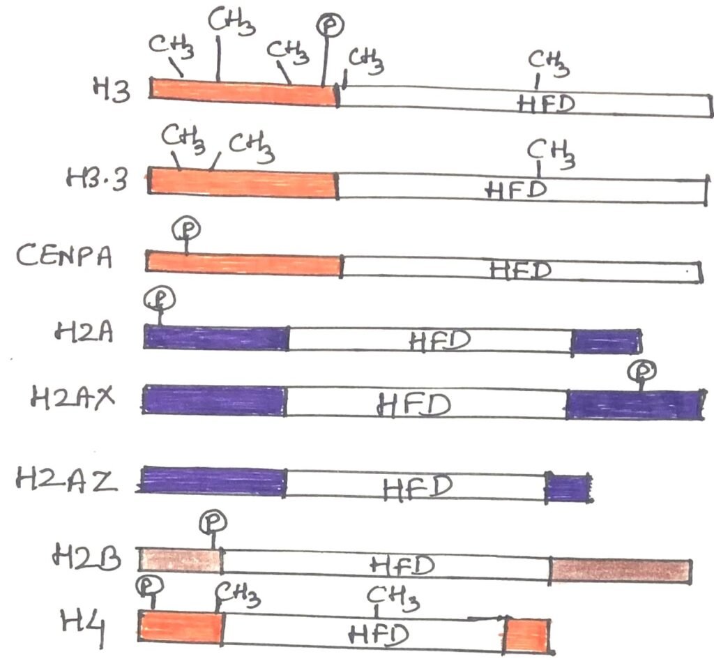
Histone H2A.Z is also incorporated into nucleosomes through specialized deposition machinery and is associated with several regulatory regions of the genome, including regions involved in transcription. Its incorporation can influence nucleosome stability and interactions between neighboring nucleosomes, thereby affecting chromatin organization and accessibility. By altering the structural properties of chromatin, H2A.Z can contribute to the regulation of gene expression. The importance of this variant is reflected in its evolutionary conservation and essential biological functions. In mammals, the gene encoding H2A.Z is essential for normal development, while in fruit flies (Drosophila), loss of H2A.Z disrupts development and prevents progression beyond the larval stages.
Histone H2A.X and DNA Damage Repair
H2A.X is a histone H2A variant that plays an important role in maintaining genome stability. It participates in the cellular response to DNA double-strand breaks. Although it is present at relatively low levels throughout the genome, it becomes particularly important when DNA damage occurs. Following a double-strand break, H2A.X molecules surrounding the damaged region are phosphorylated at serine 139 in their carboxy-terminal tails, producing a modified form known as γH2AX. This modification promotes the recruitment and assembly of proteins involved in DNA damage signaling and repair. Disruption of this phosphorylation process interferes with the formation of repair-associated protein complexes. Studies in mice have also shown that the loss of H2A.X can lead to genomic instability and male infertility. This highlights its importance in genome maintenance and reproductive function.
CENP-A and Centromere Organization
CENP-A is a specialized variant of histone H3 that replaces canonical H3 in nucleosomes at centromeres. These chromosomal regions are essential for kinetochore assembly and the accurate segregation of chromosomes during cell division. By establishing a distinctive chromatin structure, CENP-A helps specify centromere identity and supports the recruitment of proteins required for chromosome segregation. Its incorporation into centromeric chromatin depends on specialized histone assembly machinery. In mice, loss of the gene encoding CENP-A is lethal, demonstrating its essential role in normal development and chromosome function.
Studying Histone Variants Using Chromatin Immunoprecipitation
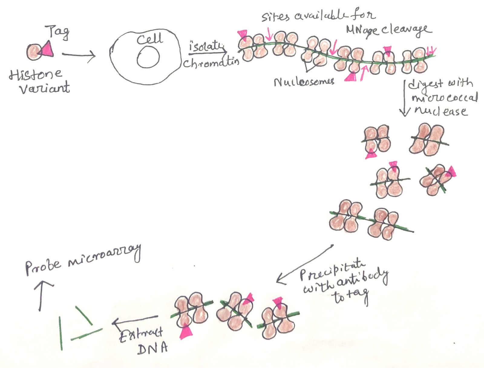
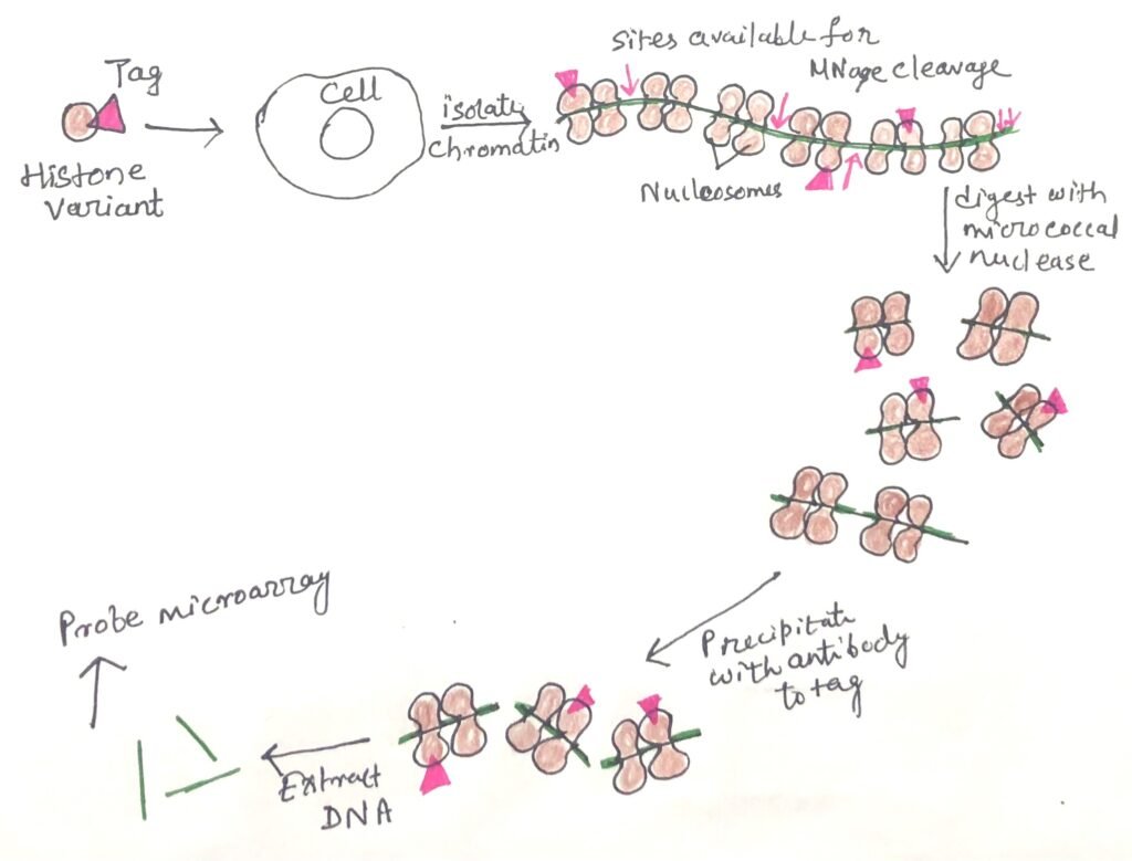
The distribution of histone variants across the genome can be investigated using chromatin immunoprecipitation (ChIP). In this technique, antibodies that specifically recognize a particular histone variant or a modified histone are used to isolate the associated chromatin fragments. The recovered material can then be analyzed to identify the DNA sequences associated with that histone variant. When combined with sequencing, a method known as ChIP-seq, this approach helps researchers map the genomic locations of histone variants and investigate their relationships with chromatin organization, gene expression, and other cellular processes.
In chromatin immunoprecipitation followed by microarray analysis (ChIP-chip), DNA recovered from immunoprecipitated chromatin is labelled and hybridized to a DNA microarray (Figure 2). This enables researchers to identify genomic regions associated with specific histone variants or histone modifications. ChIP-chip has been widely used to investigate chromatin distribution. However, sequencing-based approaches such as ChIP-seq are now commonly used for genome-wide mapping.
Histone variants and their covalent modifications contribute to the functional organization of chromatin. Depending on their identity and genomic context, they can influence processes such as transcription, DNA repair, and chromosome segregation. They do so by altering chromatin properties and recruiting specific regulatory proteins. Some histone modifications and associated chromatin states can be maintained or re-established after DNA replication and cell division, contributing to epigenetic inheritance. However, the persistence and transmission of these marks vary. Not all histone modifications are retained through cell division or meiosis.
Histone Modifications and Their Biological Significance
Histone proteins undergo several post-translational modifications that influence chromatin organization and gene activity. These chemical changes occur mainly on the histone tails and include acetylation, methylation, and phosphorylation. Histone acetylation generally promotes a more accessible chromatin environment, whereas the effects of histone methylation depend on the modified residue and the number of methyl groups added. Histone phosphorylation participates in processes such as DNA damage responses and chromosome condensation. These modifications regulate transcription, DNA repair, and other chromatin processes. Together with histone variants, they contribute to the regulation of chromatin structure and function.
Conclusion
Histone proteins provide the structural framework for DNA packaging in eukaryotic cells. Histone variants introduce specialized properties that influence chromatin organization and function. Variants such as H3.3, H2A.Z, H2A.X, and CENP-A contribute to distinct cellular processes. These processes include transcriptional regulation, DNA damage repair, and chromosome segregation. Together with covalent histone modifications, these proteins contribute to epigenetic regulation and genome stability. Understanding their distribution, deposition, and functions offers valuable insight into how chromatin structure links to cellular activity and maintains genetic information.
You may also like:

I, Swagatika Sahu (author of this website), have done my master’s in Biotechnology. I have around fourteen years of experience in writing and believe that writing is a great way to share knowledge. I hope the articles on the website will help users in enhancing their intellect in Biotechnology.