
This article describes different methods used to measure cell proliferation and division. Cell proliferation is an essential process in growth, tissue maintenance, and immune responses, making its accurate assessment important in many areas of biological research. Different experimental approaches can provide information about DNA synthesis, cellular metabolic activity, and changes in DNA content during the cell cycle. Understanding the principles and applications of these methods helps researchers select the most appropriate approach for studying cellular proliferation under different experimental conditions.
Cell Proliferation and Its Importance in Immune Responses
Cell proliferation is a fundamental biological process that is essential for growth, tissue maintenance, and the replacement of damaged cells. It also plays a crucial role in immune responses, particularly following the activation of lymphocytes by an immunological stimulus. Activated lymphocytes undergo rapid proliferation, resulting in the expansion of immune-cell populations that participate in the subsequent immune response.
The measurement of cell proliferation provides valuable information about cellular activity, immune-cell function, and cell-cycle progression. Several experimental methods have therefore been developed to assess the proliferative status of cells. These methods rely on different characteristics of dividing cells, such as DNA synthesis, metabolic activity, or changes in cellular DNA content. Commonly used approaches include tritiated thymidine uptake, colorimetric proliferation assays, bromodeoxyuridine (BrdU)-based assays, and propidium iodide staining. Although these techniques are widely used, each method has distinct principles, advantages, and limitations, making the choice of assay dependent on the experimental objectives and the type of information required.
Tritiated Thymidine Uptake Assay
Tritiated thymidine uptake was one of the earliest methods routinely used to measure lymphocyte proliferation in culture. The assay is based on the increased DNA synthesis that occurs when cells enter the proliferative phase. During this process, thymidine is incorporated into newly synthesized DNA. When radioactive thymidine, commonly tritiated thymidine, is supplied to the culture medium, actively dividing cells incorporate it into their DNA.
In this assay, lymphocytes are cultured with tritiated thymidine and exposed to an appropriate proliferative stimulus. At specific time points after stimulation, the cells are collected, and their DNA is processed for measurement. The cellular DNA containing the incorporated radioactive thymidine is retained on suitable filters, while free or unincorporated thymidine is removed during the washing steps. The radioactivity remaining on the filters is then measured. The level of radioactivity reflects the extent of DNA synthesis and therefore provides an indirect measure of lymphocyte proliferation in the culture.
Colorimetric Cell Proliferation Assays
Since tritiated thymidine is a radioactive material, its use requires appropriate radiation-safety precautions, trained personnel, and proper disposal of radioactive waste. Several colorimetric assays have been developed to estimate cell proliferation by exploiting the metabolic activity of living cells. These methods are based on the conversion of colourless chemical substrates into coloured products by cellular enzymes. The resulting colour can be quantified using spectrophotometry and provides an indirect measure of the number of metabolically active cells.
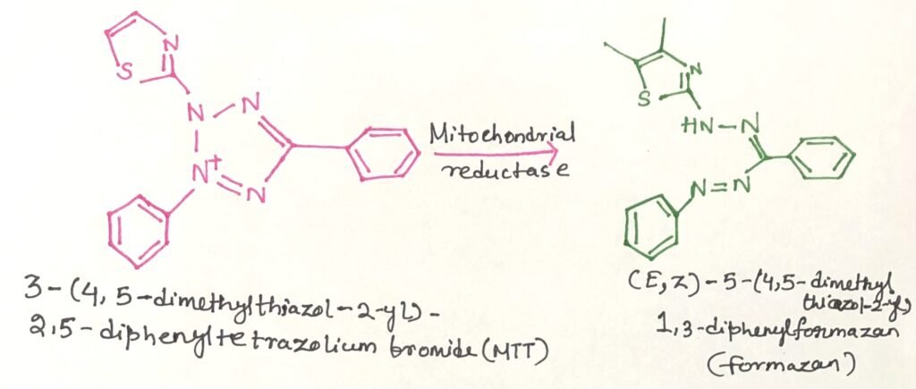
The MTT assay is one of the most widely used examples of this approach. It uses the yellow tetrazolium compound MTT, which is converted by mitochondrial NAD(P)H-dependent oxidoreductase enzymes in metabolically active cells into insoluble purple formazan crystals (Figure 1). These crystals are subsequently dissolved, and the resulting colour intensity is measured spectrophotometrically at approximately 570 nm. Under suitable experimental conditions, the amount of formazan produced correlates with the number of viable metabolically active cells. Therefore, changes in absorbance over time can be used to assess cell proliferation, while a reduction in metabolic activity can provide an indication of cell loss or death.
Bromodeoxyuridine-Based Assays
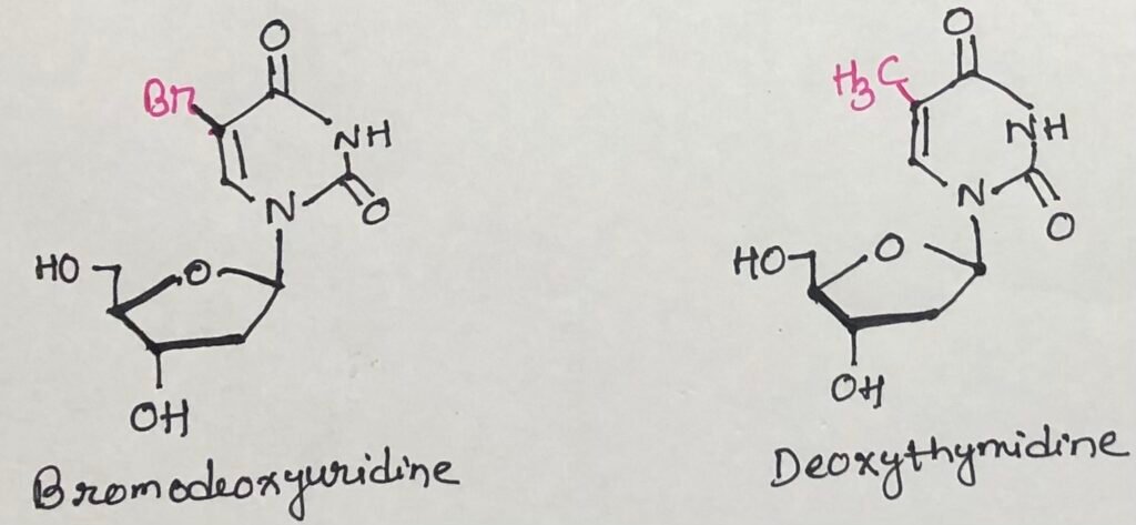
Bromodeoxyuridine (BrdU) is a synthetic analogue of deoxythymidine (Figure 2) that can be used to identify cells undergoing DNA synthesis. After entering the cell, BrdU is converted into its phosphorylated form and incorporated into newly synthesized DNA in place of thymidine. Cells containing BrdU can subsequently be detected using antibodies that specifically recognize the incorporated analogue. This provides a means of identifying cells that have entered the DNA synthesis phase and therefore helps in assessing cellular proliferation.
BrdU incorporation has also been used in approaches that selectively target recently dividing cells. Cells with BrdU levels can undergo light-induced damage when exposed to appropriate illumination, thereby selectively eliminating proliferating cell populations. In addition to BrdU, other thymidine analogues have been developed for proliferation studies. One widely used example is 5-ethynyl-2′-deoxyuridine (EdU), which can be detected through a chemical reaction between its terminal alkyne group and specific fluorescent reagents. This allows newly synthesized DNA to be visualized and provides an alternative approach for detecting proliferating cells.
Propidium Iodide-Based Cell-Cycle Analysis
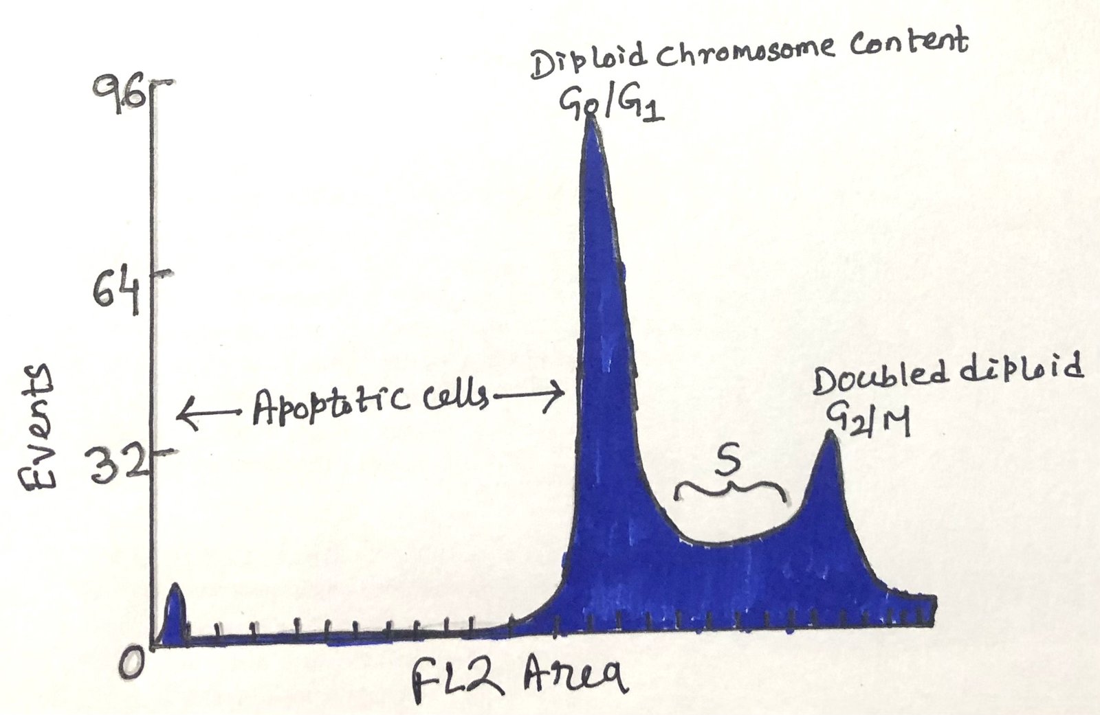
Propidium iodide (PI) is a fluorescent dye that binds to DNA and can be used to determine the DNA content of individual cells. PI fluorescence intensity reflects the DNA content of each cell. When analysed by flow cytometry, PI fluorescence is commonly detected in the red fluorescence channel. This makes it possible to distinguish cell populations according to their position within the cell cycle.
G1 cells contain about half the DNA of G2/M cells. Cells undergoing DNA synthesis in the S phase therefore show an intermediate DNA content. In addition, cells undergoing DNA degradation may display DNA levels below the normal G1 population (Figure 3). These DNA differences help estimate cells in each cell-cycle stage.
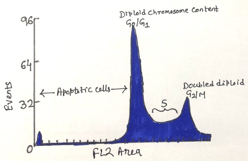
Accurate analysis also requires the exclusion of cell doublets and larger aggregates. Two attached cells may have DNA content similar to a single G2/M-phase cell. If such events are not excluded, the proportion of cells classified as being in G2/M may be artificially increased. Other DNA-binding fluorescent dyes, including DAPI, Hoechst 33342, and 7-aminoactinomycin D (7-AAD), can also be used for similar cell-cycle analyses.
Conclusion
A variety of methods are available for measuring cell proliferation and assessing cell-cycle progression. Each approach provides different types of information. Tritiated thymidine uptake measures DNA synthesis, while MTT assays assess cellular metabolic activity. BrdU and EdU identify cells undergoing DNA synthesis, while propidium iodide measures DNA content and cell-cycle distribution.
No single method is suitable for every experimental purpose. The choice of assay depends on the cell type, experimental conditions, and the specific aspect of proliferation or cell-cycle progression being investigated. Using an appropriate method, or combining complementary approaches, can provide a more reliable understanding of cellular proliferation and division. These techniques therefore remain valuable tools in immunology, cell biology, and biomedical research.
You may also like:
- Protein Kinases Regulate the Cell Cycle
- Genetic Alterations That Remove the Brakes on Cell Division
- Quantitative Cell Analysis Using Flow Cytometry

I, Swagatika Sahu (author of this website), have done my master’s in Biotechnology. I have around fourteen years of experience in writing and believe that writing is a great way to share knowledge. I hope the articles on the website will help users in enhancing their intellect in Biotechnology.