
This article focuses on the affinity between antigens and antibodies and the methods used to evaluate their binding strength. The strength of these interactions is important for understanding how antibodies recognize and bind to their specific targets. Different techniques can provide information about the extent and behavior of these molecular interactions. Accurate measurement of affinity is therefore important in immunological and biomedical research.
The Antigen-Antibody Affinity
Antigen–antibody binding is a fundamental interaction in immunology and plays an important role in many immune responses and diagnostic applications. The strength of this interaction is described by antibody affinity, which refers to the binding strength between a single antigen-binding site of an antibody and its corresponding epitope. Affinity is determined by the combined effect of multiple noncovalent forces involved in antigen–antibody binding and can be expressed quantitatively using the dissociation constant.
Several techniques are available for evaluating antigen–antibody affinity. Among these, equilibrium dialysis provides a simple and relatively inexpensive approach for studying binding interactions and demonstrating fundamental principles of affinity. Surface plasmon resonance (SPR), on the other hand, is a modern technique that enables detailed analysis of molecular binding interactions. Although SPR offers several advantages, it requires specialized instrumentation, which can increase the cost and complexity of the analysis. Understanding these methods is important for accurately characterizing the strength and behavior of antigen–antibody interactions.
Equilibrium Dialysis
Equilibrium dialysis is a classical method for determining the affinity between an antibody and its ligand. The technique uses two compartments that are separated by a semipermeable membrane. The antibody is placed in one compartment (A), while a small, labeled ligand is introduced into the other (B). The membrane allows the free ligand to pass between the two compartments but prevents the antibody from crossing it. In the absence of antibody, the ligand gradually distributes between both compartments until its concentration becomes equal on either side of the membrane (Figure 1).
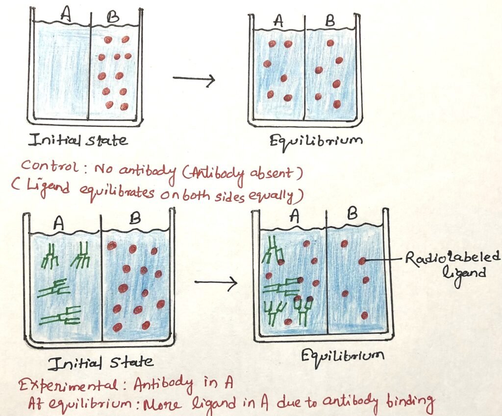
When antibody is present, some of the ligand binds to it and is retained in the antibody-containing compartment. As a result, the total ligand concentration becomes higher in the antibody compartment than in the other compartment (Figure 2). The difference between the ligand concentrations in the two compartments reflects the amount of ligand bound to the antibody.
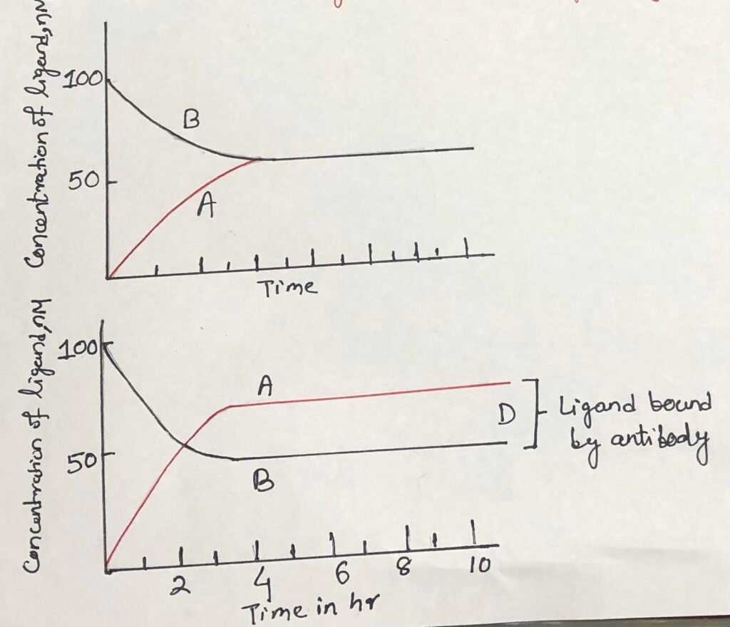
A stronger antibody–ligand interaction results in greater ligand binding and therefore a larger difference in ligand concentration between the compartments. By determining the concentrations of free and bound ligand and using the known antibody concentration, the dissociation constant (Kd) can be calculated. Thus, equilibrium dialysis provides a relatively simple way to quantify antigen–antibody affinity.
Calculation of dissociation constant (Kd)
Antibody affinity represents the strength of binding between an antibody and its corresponding antigen or epitope. This interaction results from multiple noncovalent forces between the antigen-binding site and the epitope. Affinity can be quantitatively expressed using the dissociation constant Kd, which is measured in molar units.
Kd = [S][L] / [SL]
Where:
- [S] = concentration of antigen
- [L] = concentration of free ligand
- [SL] = concentration of antigen–ligand complexes
A lower Kd value indicates stronger binding affinity, whereas a higher Kd value indicates weaker affinity.
Surface Plasmon Resonance
Surface plasmon resonance (SPR) has become an important alternative to equilibrium dialysis for determining antigen–antibody affinity. It is a rapid and sensitive technique that can provide information not only about binding affinity but also about the rates at which antigen–antibody interactions occur. This makes SPR particularly useful for studying the kinetics of molecular binding.
The technique is based on detecting changes that occur at the surface of a metal sensor when molecules bind to its surface. In a typical SPR experiment, one binding partner is immobilized on the sensor surface, while the other is introduced into the system. Binding causes changes in the optical properties at the metal–solution interface. These changes arise from surface plasmon waves, which are sensitive to alterations occurring near the metal surface. By monitoring these changes over time, the interaction between the antigen and antibody can be characterized.
Principle of Surface Plasmon Resonance
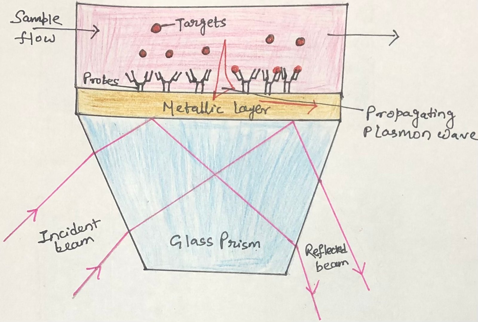
In SPR, polarized light is directed through a prism toward a sensor chip containing a thin gold film (Figure 3). The opposite surface of the film is coated with the antigen of interest. When the incident light reaches the gold layer, a portion of its energy can excite surface plasmon waves at the metal–solution interface. This produces a characteristic reduction in reflected light intensity at a specific angle, known as the resonance angle. The position of this angle depends on several factors, including the properties of the light, the gold film, and the material present near its surface.
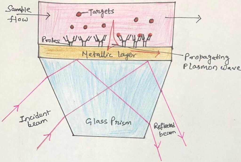
SPR detects changes in this resonance condition when antibodies bind to the immobilized antigen. The binding alters the optical properties near the gold surface, resulting in a measurable shift in the resonance angle. The magnitude of this change is related to the amount of antibody bound to the antigen. Monitoring the change over time also allows the rate of antigen–antibody binding to be determined, providing information about the kinetics of the interaction.
Operational Procedure and Kinetic Analysis
In an SPR experiment, a solution containing a known concentration of antibody is passed over a sensor surface on which the antigen has been immobilized. The interaction between the antibody and antigen produces changes in the resonance angle, which are continuously recorded over time. The resulting graph of resonance-angle changes against time is known as a sensogram. During the association phase, the signal increases as antibodies bind to the available antigen-binding sites. As these sites become occupied, the signal gradually reaches a stable level or plateau (Figure 4).
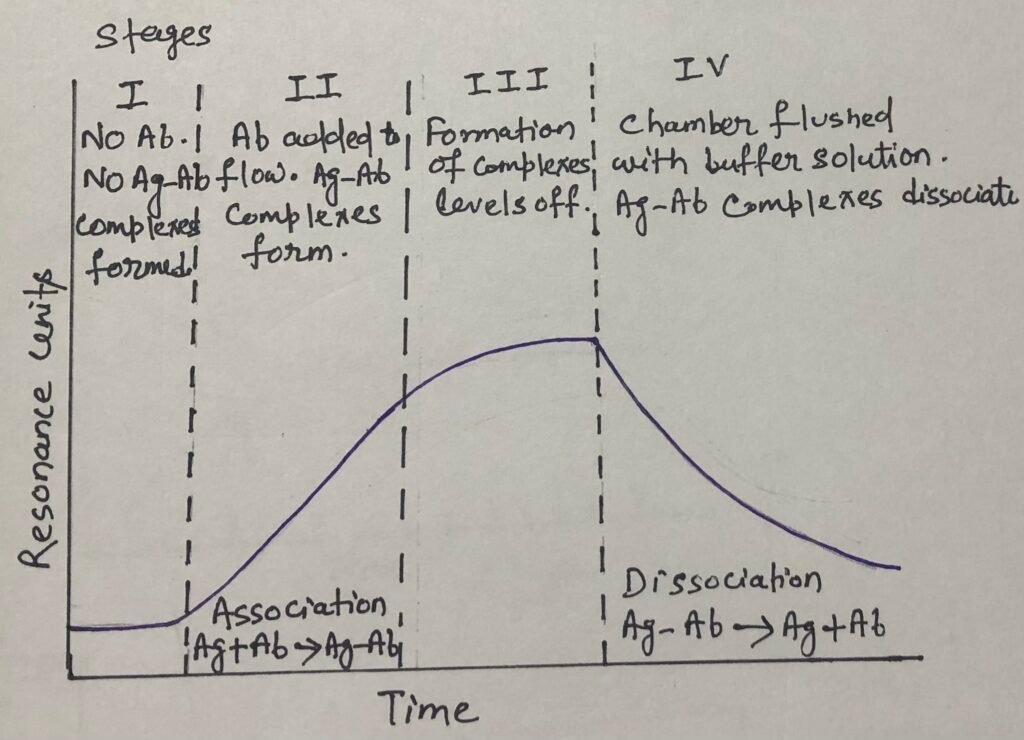
The binding data obtained during this phase can be used to determine k₁, the association rate constant, which indicates how rapidly the antibody binds to the antigen. After the binding signal reaches a plateau, the antibody-containing solution is replaced with a solution lacking antibody. The bound antibody then begins to dissociate from the antigen, causing the signal to decrease. This dissociation phase provides information for calculating k₂, the dissociation rate constant.
The values of k₁ and k₂ can subsequently be used to determine the association constant (Kₐ) according to the relationship:
Kₐ = k₁ / k₂
Thus, SPR can provide both quantitative and kinetic information about antigen–antibody interactions.
Conclusion
Determining antigen–antibody affinity is important for understanding the strength and behavior of molecular interactions. Equilibrium dialysis provides a simple approach for measuring affinity through the distribution of free and bound ligand. SPR offers a more rapid and sensitive alternative, while also providing information about the kinetics of antibody binding and dissociation. Together, these methods provide valuable approaches for characterizing antigen–antibody interactions and selecting suitable techniques for different experimental requirements.
You may also like:
- The antigen-antibody interaction- Radioimmunoassay
- The Antigen-Antibody Interaction- Immunofluorescence
- The antigen-antibody interaction: Agglutination reaction
- The antigen-antibody interaction: Precipitation reaction

I, Swagatika Sahu (author of this website), have done my master’s in Biotechnology. I have around fourteen years of experience in writing and believe that writing is a great way to share knowledge. I hope the articles on the website will help users in enhancing their intellect in Biotechnology.