
This article gives a brief description of the direct quantification of bacterial growth using cell count and cell mass. Quantitative measurement of bacterial growth is essential for studying microbial populations and their behaviour under different conditions. Various direct methods are used to estimate bacterial growth by determining either the number of cells or the total cell mass present in a culture. These techniques help scientists in microbiology, medicine, biotechnology, food analysis, and environmental studies. Methods such as microscopic counting, plate counting, membrane filtration, turbidimetric analysis, and dry weight determination each have their own advantages and limitations. This article discusses the important direct methods used for the quantitative measurement of bacterial growth.
Overview of Bacterial Growth Measurement
The growth of bacteria can be understood as the rise in their cellular population and overall biomass when favourable conditions are available. Measuring this growth quantitatively plays a major role in several scientific fields, including microbiology, biotechnology, healthcare, and food science. It helps researchers evaluate microbial multiplication and behaviour. Different techniques are used to estimate bacterial growth. However, direct measurement methods are considered more reliable since they determine the actual quantity of bacterial cells or their total mass. Two important direct approaches used for the quantitative assessment of bacterial growth are cell count and cell mass measurement.
Direct Microscopic Cell Count Method
The number of bacterial cells can be determined directly with the help of a Petroff–Hausser counting chamber. It is a specially designed microscopic slide marked with extremely small calibrated squares. Each square possesses a defined surface area, while a cover slip placed above the chamber creates a fixed and known volume for counting. Using a phase-contrast microscope, unstained bacterial cells suspended in liquid can be viewed and counted conveniently.
This technique provides a quick and straightforward way to estimate bacterial population size without requiring complex instruments. Along with counting, the physical shape and arrangement of bacterial cells can also be examined simultaneously. Highly concentrated bacterial samples may be analysed after suitable dilution to obtain an accurate count. However, the method becomes less reliable when the bacterial concentration is very low, especially during the early stages of microbial growth. This happens because very few cells are available for accurate observation and counting.
Electronic Cell Counting and Viable Cell Determination
In this technique, a bacterial sample is introduced into an electronic particle counting device. In this device, the cells pass individually through a minute opening measuring nearly 10–30 μm in diameter. The small opening links two chambers filled with an electrically conductive fluid. Whenever a bacterial cell passes through the aperture, it temporarily changes the electrical resistance between the two compartments. This change generates an electrical signal that is automatically recorded by the instrument.
This method enables rapid estimation of bacterial cell numbers and reduces manual effort in counting. However, it requires advanced electronic instruments for operation, and the narrow opening may sometimes become blocked during the process.
A major limitation of direct cell counting methods is that they cannot distinguish living cells from dead ones. To estimate the number of viable bacteria accurately, methods that permit bacterial growth and colony formation are preferred. Common approaches include the plate count method and the membrane filter technique, both of which measure only the living, actively growing cells present in the culture.
Plate Count Method for Viable Bacterial Estimation
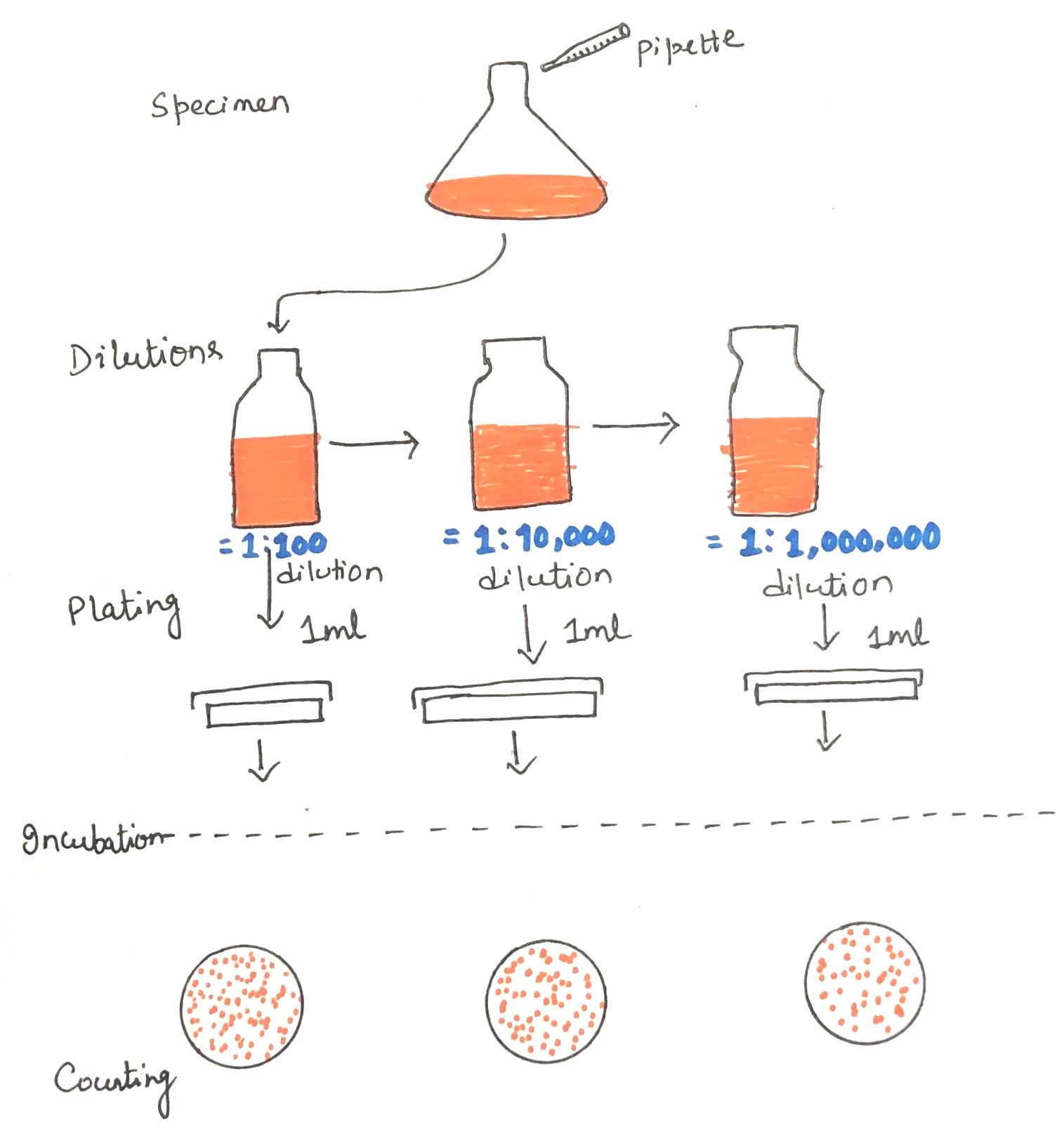
The plate count method is commonly employed to determine the number of living bacterial cells capable of reproducing under specific growth conditions. In this procedure, a known quantity of bacterial suspension is transferred into a sterile Petri plate, followed by the addition of molten agar medium. The mixture is gently rotated to distribute the microorganisms evenly throughout the medium. Once the agar solidifies, the bacterial cells become embedded within it. Each viable cell multiplies repeatedly to produce a visible cluster of cells known as a colony (Figure 1). Therefore, by counting the colonies formed, the viable microbial population present in the original sample can be estimated.
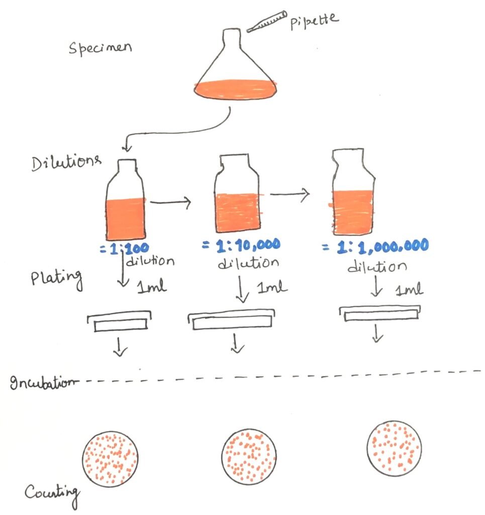
Before plating, the bacterial sample is usually diluted to obtain a manageable number of colonies, generally between 30 and 300 per plate. Counts within this range are considered reliable because overcrowding is avoided and interference among growing colonies is minimised. Colonies are often viewed using dark-field illumination, which makes them easier to distinguish. Magnifying devices may also be used for accurate counting. Modern laboratories sometimes employ electronic colony counters to simplify the process further.
Limitations of the plate count technique
Despite its usefulness, the plate count technique has certain limitations. Only bacteria that are capable of growing on the chosen culture medium will form colonies. In addition, the incubation conditions must also be suitable for their growth and detection. This becomes particularly important when analysing mixed bacterial populations. Another drawback is that a single colony may not always originate from one individual bacterial cell. Some bacteria naturally occur in groups, such as clusters, chains, or pairs, causing several cells to produce a single colony together. Consequently, the results are commonly expressed as colony-forming units per millilitre (CFU/mL) rather than the exact number of bacterial cells.
Even with these limitations, the plate count method remains one of the most widely used techniques for estimating bacterial populations in milk, water, food products, and many other materials. The method is simple, sensitive, and adaptable for detecting both small and large microbial populations accurately.
Membrane Filter Technique for Bacterial Enumeration
The membrane filter method is a modified form of the plate count technique used for estimating viable bacterial populations. In this approach, a membrane filter containing extremely small and uniform pores is used to capture microorganisms from a sample. The pore size is carefully selected so that bacterial cells are retained on the surface of the membrane while the liquid or air passes through.
This method is especially useful when analysing large samples that contain only a very small number of bacteria, such as drinking water, wastewater, or air samples. Large volumes can be processed efficiently by passing them through the filtration apparatus, allowing the microorganisms present to become concentrated on the membrane surface.
After filtration, the membrane carrying the trapped bacteria is transferred onto a culture plate or absorbent pad containing a suitable nutrient medium. During incubation, the viable bacterial cells multiply and develop into visible colonies on the membrane. Selective media and indicator dyes may also be used to improve the identification of specific groups of microorganisms. These substances make detection easier and more effective than in conventional plate count methods.
Turbidimetric Measurement of Bacterial Cell Mass
When bacterial cells are present in large numbers within a liquid suspension, they interfere with the passage of light by absorbing and scattering it. This causes the culture to appear cloudy or turbid to the naked eye. Generally, suspensions containing more than 10⁷–10⁸ cells per millilitre show noticeable turbidity. Instruments such as spectrophotometers and colorimeters are commonly used to measure this turbidity, which indirectly reflects the total bacterial cell mass in the culture (Figure 2).
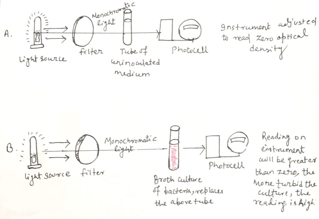
Turbidimetric analysis is widely preferred because it is quick, convenient, and suitable for continuously monitoring bacterial growth. However, the method is effective only when the bacterial population is sufficiently dense to produce measurable cloudiness. Accurate readings may become difficult if the growth medium itself is strongly colored. Errors may also arise when the suspension contains particulate materials other than bacteria. In addition, turbidity measurements do not distinguish between living and dead cells, since both contribute equally to the cloudiness of the culture.
Measurement of Bacterial Growth by Nitrogen Estimation
Proteins form a major portion of bacterial cell material. Since nitrogen is an essential component of proteins, the amount of nitrogen present can be used to estimate bacterial growth. On a dry-weight basis, bacterial cells generally contain about 14% nitrogen. Therefore, determining the nitrogen content of a culture provides an indirect measurement of the bacterial biomass.
In this method, bacterial cells are first collected from the culture and carefully washed to remove any traces of the surrounding medium. After purification, the sample undergoes a quantitative chemical analysis to determine its nitrogen content. The measured nitrogen level is then used to estimate the size of the bacterial population.
Although this technique can provide reliable results, it is relatively time-consuming and requires considerable laboratory effort. The method can only be applied to samples that are completely free from external nitrogen-containing substances, as contamination may interfere with accurate measurements. In addition, it is suitable mainly for dense bacterial populations rather than dilute cultures. Due to these limitations, nitrogen estimation is used mostly in specialised microbiological research rather than routine laboratory analysis.
Determination of Bacterial Growth by Dry Weight Measurement
Dry weight estimation is considered one of the most direct methods for measuring the total mass of bacterial cells in a culture. In this technique, bacterial cells are collected, thoroughly washed to eliminate impurities and residual medium components. Then the cells are dried before their mass is determined. The measured dry weight reflects the overall quantity of cellular material present in the sample.
Despite its direct nature, this method is suitable only for highly concentrated bacterial suspensions. Because small populations do not provide enough material for accurate measurement. In addition, all non-cellular substances must be removed carefully to avoid errors in the final weight determination.
Another important limitation is that an increase in dry weight does not always correspond to an actual rise in living cell numbers. Certain bacteria may accumulate intracellular storage compounds during later stages of growth. For instance, Azotobacter beijerinckii can store large amounts of poly-β-hydroxybutyrate during the stationary phase. This reserve material may constitute a major portion of the cell’s dry mass. As a result, the dry weight of the culture may continue to rise even when true bacterial growth has slowed or stopped.
Conclusion
Quantitative measurement of bacterial growth is essential for understanding microbial populations and their activities. Direct methods such as cell counting and cell mass determination provide reliable ways to estimate bacterial growth under laboratory conditions. Although each method has its own advantages and limitations, together they play an important role in microbiological research, medical studies, food analysis, and biotechnology.
You may also like:
- The green phototrophic bacteria
- Biochemical and Physiological Characterization of Milk Bacteria
- Coliform Bacteria as Indicators of Water Quality


I, Swagatika Sahu (author of this website), have done my master’s in Biotechnology. I have around fourteen years of experience in writing and believe that writing is a great way to share knowledge. I hope the articles on the website will help users in enhancing their intellect in Biotechnology.