
In this article, I briefly explain clonal anergy and its relation with a costimulatory signal.
Clonal anergy
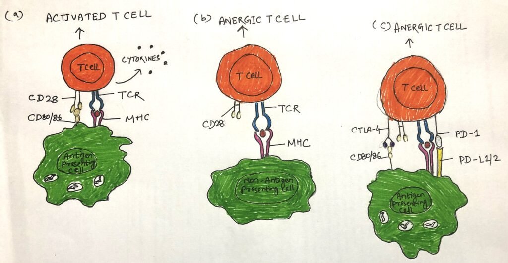
A state of clonal anergy develops, when a naïve T cell engages its TCR with an antigen presented by an MHC, without a suitable costimulatory signal. It is a state, where the specific T cell clone shows no response to subsequent stimulation.
T cells specific for an MHC-peptide complex can be induced to proliferate in vitro by incubation with activated antigen-presenting cells expressing both MHC peptide complex and CD80/86.
When T cells are exposed to fixed antigen-presenting cells that can not up-regulate CD80/86, make T cells unresponsive. These T cells become anergic (Figure 1b) and unable to secrete cytokines or proliferate even in response to subsequent stimulation.
The conditions that develop a state of anergy
A costimulatory signal inhibits the activation of auto-reactive T cells
A T cell gets activated by a costimulatory signal. Without this, auto-reactive T cells that have escaped the thymus will be activated and pose a threat.
A naïve T cell with a T cell receptor, specific for an MHC class-I insulin peptide complex, would become non-responsive when it encounters a pancreatic islet beta cell expressing an MHC class-I peptide complex. This happens as islet beta cells can’t be induced to express costimulatory ligands. Thus, this results in a state of anergy.
Interactions between coinhibitory receptors and ligands induce anergy
Activated T cells that have upregulated coinhibitory receptors, could aid in restraining proliferation of T cells after clearance of antigen. During chronic infection caused by mycobacteria, HIV, hepatitis virus, and exposure to tumor antigens, coinhibition may bring about T cell exhaustion. T cells, specific for these pathogens and antigens express elevated levels of coinhibitory receptors such as PD-1 and BTLA. Thus, the T cells become functionally anergic (figure 1C). To block such coinhibitory interactions, recent therapies include the involvement of checkpoint antibodies that work magically in cancer therapy.
Signal 3: Required for T cell activation
When a naïve T cell comes in contact with an antigen-presenting cell, it gets activated by the engagement of its TCR with the antigen-MHC complex. The costimulatory receptors also aid in the activation, by engaging themselves with the specific costimulatory ligands. However, the activity of soluble cytokines, produced by antigen-presenting cells and T cells, also plays an important role in activating T cells.
IL-2 is one of the vital cytokines involved in T cell activation. TCR and costimulatory signals induce the transcription of genes for IL-2 and high-affinity IL-2 receptors. These signals also strengthen the stability of IL-2 mRNA. Activated T cell increases IL-2 production by a hundredfold. The binding of IL-2 and the high affinity IL-2 receptor spur activated naïve T cells for rapid proliferation.
A variety of cell types including antigen-presenting cells (APCs), T cells, and innate lymphoid cells produce an important set of cytokines, known as polarizing cytokines. These cytokines play a key role in determining the type of effector cell that a T cell becomes.
Conclusion
Clonal anergy is a state, where the specific T cell clone shows no response to subsequent stimulation. When T cells are exposed to fixed antigen-presenting cells that can not up-regulate CD80/86, make T cells unresponsive. These T cells become anergic and unable to secrete cytokines or proliferate even in response to subsequent stimulation.

During chronic infection caused by mycobacteria, HIV, hepatitis virus, and exposure to tumor antigens, coinhibition may bring about T cell exhaustion. T cells, specific for these pathogens and antigens express elevated levels of coinhibitory receptors such as PD-1 and BTLA.
When a naïve T cell comes in contact with an antigen-presenting cell, it gets activated by the engagement of its TCR with the antigen-MHC complex. The costimulatory receptors also aid in the activation, by engaging themselves with the specific costimulatory ligands.
You may also like:

I, Swagatika Sahu (author of this website), have done my master’s in Biotechnology. I have around fourteen years of experience in writing and believe that writing is a great way to share knowledge. I hope the articles on the website will help users in enhancing their intellect in Biotechnology.